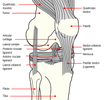
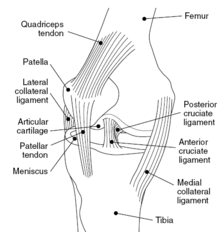
Knee
The knee joint joins the thigh with the leg and consists of two articulations: one between the femur and tibia, and one between the femur and patella. It is the largest joint in the human body.The knee is a mobile trocho-ginglymus(a hinge joint), which permits flexion and extension as well as slight internal and external rotation. The knee joint is vulnerable to both acute injury and the development of osteoarthritis.
It is often grouped into tibiofemoral and patellofemoral components. (Thefibular collateral ligament is often considered with tibiofemoral components.)
Structure
The knee is a hinge type synovial joint, which is composed of three functional compartments: the femoropatellar articulation, consisting of the patella, or "kneecap", and the patellar groove on the front of the femur through which it slides; and the medial and lateral femorotibial articulations linking the femur, or thigh bone, with the tibia, the main bone of the lower leg. The joint is bathed in synovial fluidwhich is contained inside the synovial membrane called the joint capsule. Theposterolateral corner of the knee is an area that has recently been the subject of renewed scrutiny and research.
The knee is one of the most important joints of our body. It plays an essential role in movement related to carrying the body weight in horizontal (running and walking) and vertical (jumps) directions.
At birth, a baby will not have a conventional knee cap, but a growth formed of cartilage. By the time that the child is 3–5 years of age, ossification will have replaced the cartilage with bone. Because it is the largest sesamoid bone in the human body, the ossification process takes significantly longer.
Articular bodies
The articular bodies of the femur are its lateral and medial condyles. These diverge slightly distally and posteriorly, with the lateral condyle being wider in front than at the back while the medial condyle is of more constant width. The radius of the condyles' curvature in the sagittal planebecomes smaller toward the back. This diminishing radius produces a series of involute midpoints (i.e. located on a spiral). The resulting series of transverse axes permit the sliding and rolling motion in the flexing knee while ensuring the collateral ligaments are sufficiently lax to permit the rotation associated with the curvature of the medial condyle about a vertical axis.
The pair of tibial condyles are separated by the intercondylar eminence composed of a lateral and a medial tubercle.
The patella is inserted into the thin anterior wall of the joint capsule. On its posterior surface is a lateral and a medial articular surface, both of which communicate with the patellar surface which unites the two femoral condyles on the anterior side of the bone's distal end.
Articular capsule
Main article: Articular capsule of the knee joint
The articular capsule has a synovial and a fibrous membrane separated by fatty deposits. Anteriorly, the synovial membrane is attached on the margin of the cartilage both on the femur and the tibia, but on the femur, the suprapatellar bursa or recess extends the joint space proximally. The suprapatellar bursa is prevented from being pinched during extension by the articularis genus muscle. Behind, the synovial membrane is attached to the margins of the two femoral condyles which produces two extensions similar to the anterior recess. Between these two extensions, the synovial membrane passes in front of the two cruciate ligaments at the center of the joint, thus forming a pocket direct inward.
Bursae
Main article: Bursae of the knee joint
Numerous bursae surround the knee joint. The largest communicative bursa is the suprapatellar bursa described above. Four considerably smaller bursae are located on the back of the knee. Two non-communicative bursae are located in front of the patella and below the patellar tendon, and others are sometimes present.
Cartilage
Cartilage is a thin, elastic tissue that protects the bone and makes certain that the joint surfaces can slide easily over each other. Cartilage ensures supple knee movement. There are two types of joint cartilage in the knees: fibrous cartilage (themeniscus) and hyaline cartilage. Fibrous cartilage has tensile strength and can resist pressure. Hyaline cartilage covers the surface along which the joints move. Cartilage will wear over the years. Cartilage has a very limited capacity for self-restoration. The newly formed tissue will generally consist of a large part of fibrous cartilage of lesser quality than the original hyaline cartilage. As a result, new cracks and tears will form in the cartilage over time.
Menisci
The articular disks of the knee-joint are called menisci because they only partly divide the joint space. These two disks, the medial meniscus and the lateral meniscus, consist of connective tissue with extensive collagen fibers containing cartilage-like cells. Strong fibers run along the menisci from one attachment to the other, while weaker radial fibers are interlaced with the former. The menisci are flattened at the center of the knee joint, fused with the synovial membrane laterally, and can move over the tibial surface.
The menisci serve to protect the ends of the bones from rubbing on each other and to effectively deepen the tibial sockets into which the femur attaches. They also play a role in shock absorption, and may be cracked, or torn, when the knee is forcefully rotated and/or bent.
Ligaments
The ligaments surrounding the knee joint offer stability by limiting movements and, together with several menisci and bursae, protect the articular capsule.
Intracapsular
The knee is stabilized by a pair of cruciate ligaments. The anterior cruciate ligament(ACL) stretches from the lateral condyle of femur to the anterior intercondylar area. The ACL is critically important because it prevents the tibia from being pushed too far anterior relative to the femur. It is often torn during twisting or bending of the knee. The posterior cruciate ligament (PCL) stretches from medial condyle of femurto the posterior intercondylar area. Injury to this ligament is uncommon but can occur as a direct result of forced trauma to the ligament. This ligament prevents posterior displacement of the tibia relative to the femur.
The transverse ligament stretches from the lateral meniscus to the medial meniscus. It passes in front of the menisci. It is divided into several strips in 10% of cases. The two menisci are attached to each other anteriorly by the ligament.The posterior and anterior meniscofemoral ligaments stretch from the posterior horn of the lateral meniscus to the medial femoral condyle. They pass posteriorly behind the posterior cruciate ligament. The posterior meniscofemoral ligament is more commonly present (30%); both ligaments are present less often. Themeniscotibial ligaments (or "coronary") stretches from inferior edges of the mensici to the periphery of the tibial plateaus.
Extracapsular
The patellar ligament connects the patella to the tuberosity of the tibia. It is also occasionally called the patellar tendon because there is no definite separation between the quadriceps tendon (which surrounds the patella) and the area connecting the patella to the tibia. This very strong ligament helps give the patella its mechanical leverage and also functions as a cap for the condyles of the femur. Laterally and medially to the patellar ligament the lateral and medial patellar retinacula connect fibers from the vasti lateralisand medialis muscles to the tibia. Some fibers from the iliotibial tract radiate into the lateral retinaculum and the medial retinaculum receives some transverse fibers arising on the medial femoral epicondyle.
The medial collateral ligament (MCL a.k.a. "tibial") stretches from the medial epicondyle of the femur to the medial tibial condyle. It is composed of three groups of fibers, one stretching between the two bones, and two fused with the medial meniscus. The MCL is partly covered by the pes anserinus and the tendon of the semimembranosus passes under it. It protects the medial side of the knee from being bent open by a stress applied to the lateral side of the knee (a valgusforce). The lateral collateral ligament (LCL a.k.a. "fibular") stretches from the lateral epicondyle of the femur to the head of fibula. It is separate from both the joint capsule and the lateral meniscus. It protects the lateral side from an inside bending force (a varus force). The anterolateral ligament (ALL) is situated in front of the LCL.
Lastly, there are two ligaments on the dorsal side of the knee. The oblique popliteal ligament is a radiation of the tendon of the semimembranosus on the medial side, from where it is direct laterally and proximally. The arcuate popliteal ligamentoriginates on the apex of the head of the fibula to stretch proximally, crosses the tendon of the popliteus muscle, and passes into the capsule.


No comments:
Post a Comment