Kidney
The kidneys are bean-shaped organs that serve several essential regulatory roles in vertebrates. They remove excess organic molecules from the blood, and it is by this action that their best-known function is performed: the removal of waste products of metabolism. Kidneys are essential to the urinary systemand also serve homeostatic functions such as the regulation of electrolytes, maintenance of acid–base balance, and regulation of blood pressure (via maintaining the salt and water balance). They serve the body as a natural filter of the blood, and remove water-soluble wastes which are diverted to thebladder. In producing urine, the kidneys excrete wastes such as urea andammonium. They are also responsible for the reabsorption of water, glucose, and amino acids. The kidneys also produce hormones including calcitriol,erythropoietin, and the enzyme renin, the last of which indirectly acts on the kidney in negative feedback.
Located at the rear of the abdominal cavity in the retroperitoneal space, the kidneys receive blood from the paired renal arteries, and drain into the pairedrenal veins. Each kidney excretes urine into a ureter which empties into the bladder.
Renal physiology is the study of kidney function, while nephrology is the medical specialty concerned with kidney diseases. Diseases of the kidney are diverse, but individuals with kidney disease frequently display characteristic clinical features. Common clinical conditions involving the kidney include thenephritic and nephrotic syndromes, renal cysts, acute kidney injury, chronic kidney disease, urinary tract infection, nephrolithiasis, and urinary tract obstruction. Various cancers of the kidney exist. The most common adult renal cancer is renal cell carcinoma. Cancers, cysts, and some other renal conditions can be managed with removal of the kidney. This is known asnephrectomy. When renal function, measured by the glomerular filtration rate, is persistently poor, dialysis and kidney transplantation may be treatment options. Although they are not normally harmful, kidney stone can be painful.
Structure
Location
In humans, the kidneys are located in the abdominal cavity, one on each side of the spine, and lie in a retroperitoneal position at a slightly oblique angle. The asymmetry within the abdominal cavity, caused by the position of the liver, typically results in the right kidney being slightly lower and smaller than the left, and being placed slightly more to the middle than the left kidney. The left kidney is approximately at the vertebral level T12 to L3, and the right is slightly lower. The right kidney sits just below the diaphragm and posterior to the liver. The left sits below the diaphragm and posterior to the spleen. On top of each kidney is an adrenal gland. The upper parts of the kidneys are partially protected by the 11th and 12th ribs. Each kidney, with its adrenal gland, is surrounded by two layers of fat: the perirenal and pararenalfat) and the renal fascia. In adult males, the kidney weighs between 125 and 170 grams. In females the weight of the kidney is between 115 and 155 grams.
Structure
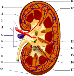
The kidney has a bean-shaped structure having a convex and a concaveborder. A recessed area on the concave border is the renal hilum, where therenal artery enters the kidney and the renal vein and ureter leave. The kidney is surrounded by tough fibrous tissue, the renal capsule, which is itself surrounded by perirenal fat (adipose capsule), renal fascia, and pararenal fat (paranephric body). The anterior (front) surface of these tissues is theperitoneum, while the posterior (rear) surface is the transversalis fascia.
The superior pole of the right kidney is adjacent to the liver. For the left kidney, it's next to the spleen. Both, therefore, move down upon inhalation.
The kidney is approximately 11–14 cm (4.3–5.5 in) in length, 6 cm (2.4 in) wide and 4 cm (1.6 in) thick.
The substance, or parenchyma, of the kidney is divided into two major structures: the outer renal cortex and the inner renal medulla. Grossly, these structures take the shape of eight to 18 cone-shaped renal lobes, each containing renal cortex surrounding a portion of medulla called a renal pyramid(of Malpighi). Between the renal pyramids are projections of cortex calledrenal columns (or Bertin columns). Nephrons, the urine-producing functional structures of the kidney, span the cortex and medulla. The initial filtering portion of a nephron is the renal corpuscle which is located in the cortex. This is followed by a renal tubule that passes from the cortex deep into the medullary pyramids. Part of the renal cortex, amedullary ray is a collection of renal tubules that drain into a single collecting duct.
The tip, or papilla, of each pyramid empties urine into a minor calyx; minor calyces empty into major calyces, and major calyces empty into the renal pelvis. This becomes the ureter. At the hilum, the ureter and renal vein exit the kidney and the renal artery enters. Hilar fat snd lymphatic tissue with lymph nodes surrounds these structures. The hilar fat is contiguous with a fat-filled cavity called the renal sinus. The renal sinus collectively contains the renal pelvis and calyces and separates these structures from the renal medullary tissue.
Blood supply
The renal circulation supplies the blood to the kidneys via the renal arteries, left and right, which branch directly from the abdominal aorta. Despite their relatively small size, the kidneys receive approximately 20% of the cardiac output.[7]
Each renal artery branches into segmental arteries, dividing further into interlobar arteries, which penetrate the renal capsule and extend through the renal columns between the renal pyramids. The interlobar arteries then supply blood to thearcuate arteries that run through the boundary of the cortex and the medulla. Each arcuate artery supplies several interlobular arteries that feed into the afferent arteriole that supply the glomeruli.
The medullary interstitium is the functional space in the kidney beneath the individual filters (glomeruli), which are rich in blood vessels. The interstitium absorbs fluid recovered from urine. Various conditions can lead to scarring and congestion of this area, which can cause kidney dysfunction and failure.
After filtration occurs, the blood moves through a small network of venules that converge into interlobular veins. As with the arteriole distribution, the veins follow the same pattern: the interlobular provide blood to the arcuate veins then back to the interlobar veins, which come to form the renal vein exiting the kidney for transfusion for blood.
Histology
Renal histology studies the microscopic structure of the kidney. Distinct cell typesinclude:
- Kidney glomerulus parietal cell
- Kidney glomerulus podocyte
- Kidney proximal tubule brush border cell
- Loop of Henle thin segment cell
- Thick ascending limb cell
- Kidney distal tubule cell
- Collecting duct principal cell
- Collecting duct intercalated cell
- Interstitial kidney cells
The renal artery enters into the kidney at the level of the first lumbar vertebra just below the superior mesenteric artery. As it enters the kidney, it divides into branches: first the segmental artery, which divides into 2 or 3 lobar arteries, then further divides into interlobar arteries, which further divide into the arcuate artery, which leads into the interlobular artery, which form afferent arterioles. The afferent arterioles form the glomerulus (network of capillaries enclosed in Bowman's capsule). From here, efferent arterioles leaves the glomerulus and divide into peritubular capillaries, which drain into the interlobular veins and then into arcuate vein and then into interlobar vein, which runs into lobar vein, which opens into the segmental vein and which drains into the renal vein, and then from it blood moves into the inferior vena cava.
Innervation
The kidney and nervous system communicate via the renal plexus, whose fibers course along the renal arteries to reach each kidney Input from the sympathetic nervous system triggers vasoconstriction in the kidney, thereby reducing renal blood flow. The kidney also receives input from the parasympathetic nervous system, by way of the renal branches of the vagus nerve (cranial nerve X); the function of this is yet unclear. Sensory input from the kidney travels to the T10-11 levels of the spinal cord and is sensed in the corresponding dermatome. Thus, pain in the flank region may be referred from corresponding kidney.
Development
Main article: Kidney development
The mammalian kidney develops from intermediate mesoderm. Kidney development, also called nephrogenesis, proceeds through a series of three successive developmental phases: the pronephros, mesonephros, and metanephros.


No comments:
Post a Comment